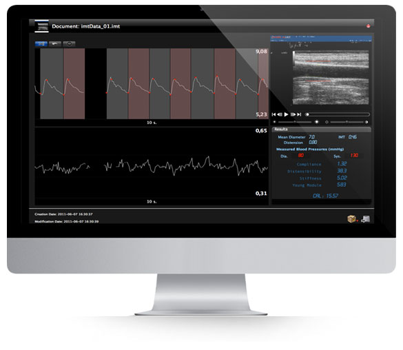
We invite you to download a 14-day fully functional trial version of Cardiovascular Suite, including FMD Studio and Carotid Studio. The software runs on Apple and Windows Computers, activation by email and Internet connection are required.
Carotid Studio
Carotid Stiffness and Intima Media Thickness
Simultaneous measurement
Echotracking precision
Easy editing
Real-time analysis
Today it is important to fully characterize the health of the carotid arteries. With our software you can have both the thickness and the stiffness of the artery from the same ultrasound scan.

Trial Version
Ver 4.6.1 (build 105) Jul 11, 2023
Minimum system requirements:
APPLE COMPUTER
Apple Mac Computer with: Intel Core i5 5th generation 2.3 GHz Turbo boost, 4GB RAM, 1GB free Hard Disk space*, USB 3.0 port, 1280x800 monitor resolution, Mac OS X 10.12 - 10.15.
MICROSOFT WINDOWS COMPUTER
Intel Core i5 5th generation 2.3 GHz Turbo boost, 4GB RAM, 1GB free Hard Disk space*, USB 3.0 port, 1024x768 monitor resolution.
Microsoft Windows 7 64 bit, Windows 8.1 64 bit, Windows 10 64 bit, OpenGL ES 2.1.
(*) 250GB free Hard Disk space is suggested for the Archive
Suggested computer:
APPLE COMPUTER
MacBook Air o MacBook Pro, processor M2 or later, 8-16GB RAM, 512GB SSD (larger for more than 1000 FMD studies), Display 13’’-16’’, macOS 13 or superior.
MICROSOFT WINDOWS COMPUTER
Laptop with Intel Core i5-i7 12000+ or AMD Ryzen 5-7 5000+, 16GB RAM, 512GB SSD (larger for more than 1000 FMD studies), Display 14’’-16’’, resolution 1920x1080, Windows 10 or 11.
To receive the download link of the 14-days trial version of Cardiovascular Suite, please enter the information requested below. We will send you an email with the details of the trial version and the link to download the software.
Features
Simultaneous measurement
Monitoring of the parameters of thickness and stiffness of the artery with assessment of the elastic properties of the arterial wall material.
Echotracking precision
Easy editing
Real-time analysis

Carotid Stiffness and Intima Media Thickness
In recent years, great emphasis has been placed on the role of arterial stiffness in the development of cardiovascular disease. Indeed, arterial stiffness assessment is increasingly used in the clinical assessment of patients. Local measurements on superficial arteries (mainly the carotid artery) can be performed using ultrasound. This approach provides optimal conditions for a precise determination of arterial stiffness, because in this way the measurement is directly obtained from the change in pressure driving the change in volume, i.e., without using any mathematical model of the circulation.
Ultrasound imaging can also provide the value of the Carotid Intima Media Thickness (IMT), a measure of the thickness of the artery. This measurement is now widely used and well-accepted as an index of atherosclerosis. In addition, the stiffness and thickness of the artery can be combined to provide a value for the elastic properties of the arterial wall material (Young's elastic module).
Validation
Carotid Studio has been validated in 87 healthy subjects (mean age 48 ± 14 years) and 311 patients with more than one cardiovascular risk factor (mean age 58 ± 11 years). Carotid Intima Media Thickness was greater in patients than in healthy subjects (0.75 ± 0.26 mm vs 0.68±0.15 mm, p = 0.001),
Carotid studio has been compared with echo-tracking technology to evaluate the accuracy of carotid intima media thickness (IMT), diameter (D) and stroke changes in diameter (ΔD) measures. Bland Altman analysis showed a bias ± standard deviation of 0.006 ± 0.039mm for IMT, 0.060 ± 0.110mm for D and 0.016 ± 0.039mm for ΔD. In addition, reproducibility was evaluated and intra-observer CVs were 6 ± 6% for IMT, 2 ± 1% for D and 8 ± 6% for IMT. Results showed that Carotid Studio overcomes the limitations of standard image analysis and that it is a suitable device for measuring IMT and local arterial stiffness parameters in clinical studies.
Carotid studio is one of the automatic measurement systems considered by the European Network for Non-Invasive Investigation of Large Arteries.

